
Sperm is the male sexual cell that is eliminated at the vagina level during intercourse.
It’s made up of head, body, and tail that gives it mobility.
A number of 200 million sperm is eliminated, but only 200 manage to pass the vagina, uterus and then the fallopian tubes to reach the oocyte. It is known that a sperm can traverse this route even in 5 minutes, but the average is 20 minutes. Their advancement is facilitated by both cervical mucus, striațiile fallopian uterine and uterus movements. The egg retains its capacity to be fertilized for approximately 12-24 hours, but sperm has a longer life in the internal environment, approximately 48-72 hours.
The oocyte membrane allows the penetration of the sperm from the contact with enzymes that are released from the level of the sperm head; When one manages to penetrate at the oocyte level and produce fertilization, the membrane becomes impenetrable to the rest, thus preventing the same oocyte from being fertilized by several sperm and the occurrence of an abnormal number of chromosomes. After the penetration of the sperm head containing the genetic material (23 chromosomes), it will unite with the genetic material of oocyte (23 chromosomes) and will result in a cell with a full number of chromosomes (46) which, by successive divisions will give rise to the egg , embryo and fetus. Fecundația takes place at the fallopian level and the egg is still moving from the first hours to the uterine cavity where it will arrive on day 6-7 and will be implanted. Abnormal implantation at the uterine fallopian level will give rise to the ectopic pregnancy.
Under the influence of estrogens and progesterone secreted by the cells of the ovarian follicles, the endometrium (lining of the uterus) is prepared for the nidation process (attachment of the egg to the uterine lining).
The placenta starts formation on day 7-8 after the fertilisation of the oocyte, having 4 stages of development:
- Postconception stage (days 7-8),
- Lacunar (days 9-12),
- Elaboration (day 13-month IV at the end)
- Status period (end of month IV-term).
The placenta develops during this period around the embryo, helping in the functions of:
- Digestion
- Excretion
- Breathing
- Endocrine role
- Protection for the conception product.
The circulatory system is inverted to the fetus; thus, CO2 is transported via the umbilical artery to the maternal circulation and the intake of O2 and nutrients to the foetus is ensured via umbilical vein. A number of hormones are also synthesized in the placenta, which can be considered a temporary endocrine organ.
Since the first trimester the fetus is able of even active movements that are not perceived by the mother than around week 20 of amenorrhea (from the last menstruation). The main organs appear, and these will develop in the next period, some such as liver, brain, kidney and lungs, functioning even in an adaptive form. During this period, the heart starts to beat with a frequency of 100-150 beats/minute.
In addition to the heart and primary organs, it develops a primary outline of the face with the already visible nose and eyelids. Other elements such as lips and ear pavilions begin to viewable in the first trimester. The kidneys are functional and produce urine that is eliminated in the amniotic fluid. The skin is still transparent. He weighs at the end of the first quarter around 23 grams and has a length of 6.7 cm.
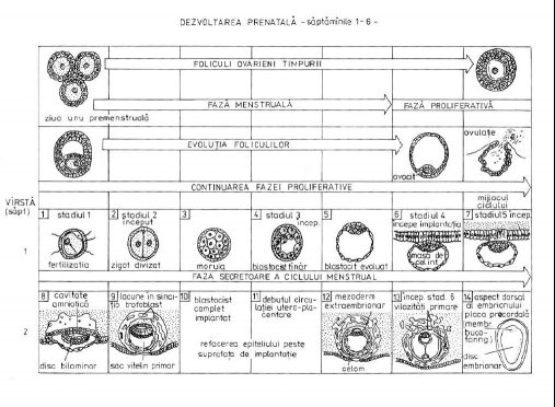
Prenatal development-Weeks 1 to 6 [Sursa, p313]
In the second trimester of pregnancy, the fetus develops the genital apparatus at the beginning of this period, and the sex of the conception product can be observed in ultrasound.
The girls appear vagina with the uterus. The fetus develops strong reflexes; it starts to sigh, but without making a sound because it has the airways filled with fluid, this reflex act produces as preparation for the breathing process.
At the end of these weeks, the fetus is well proportionted, covered with hair and relatively opaque skin, starting to look more and better with a newborn. It develops a form of hearing being able to listen to the distorted voice of the mother and react to various sounds, frequencies and a certain level of intensity. It opens its eyes, dreams and sleeps with its own sleep-waking rhythm. It weighs 875 gr in these moments and has a size of 36.6 cm.
In the first part of the 3rd quarter, the last trimester before birth, the child develops its eyes being able to perceive the luminous stimuli that penetrate the mother’s belly. The fetus begins to absorb the necessary minerals such as iron and calcium, defending even its fingernails. As for the sexual characters, they are at an exponential stage of evolution, the testicles being lowered into the scrotum and the clitoris, not being initially covered by the labia large, is prominently visible.
In the week 33 of gestation, in most cases the fetus will be positioned with the cephalic extremity at the lower uterine pole, being prepared for childbirth. Throughout this period, the lungs, heart and nervous system are in continuous development, along with the fat tissue of the fetus, which has a role in thermo-adjustment after birth. With fully functional kidneys and liver almost completely developed alongside them and most of the main systems that have reached the final stage of development, the child takes very fast weight in the last 3 weeks.
In week 39, the fetus is perfectly developed, with an average weight of about 3 kg and 49.5 cm.
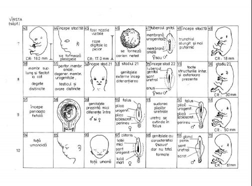
Development of the fetus-weeks 7 to 10 [Sursa, p314]

